Veterinary Urgent Care by ETHOS

Diagnostic Equipment
Diagnostic Equipment
Radiographs
Commonly referred to as X-rays, radiographs diagnose diseases and conditions in the chest, abdomen, and musculoskeletal system (this includes bones, muscles, tendons, ligaments, and soft tissue). Radiographs are the most common diagnostic tool used in veterinary medicine. They are fast and accurate at diagnosing a wide variety of conditions, including:
Foreign bodies – Gastrointestinal obstruction
Urinary obstructions – If you think your pet might have a urinary obstruction, please head directly to a 24/7 emergency room. This can be a life-threatening emergency. Urinary blockages more commonly affect cats and are especially dangerous in male cats.
Bone fractures and other trauma
Respiratory distress and pneumonia
Pregnancy
X-rays are safe, non-invasive, and do not cause discomfort. In some cases sedation may be recommended to relieve signs of anxiety or stress your pet may exhibit during the procedure.
Comprehensive Lab Equipment
Veterinary Urgent Care clinics have in-house comprehensive lab equipment to run full bloodwork and urinalysis on your pet. Our clinics also use a larger reference lab for more in-depth tests that can be sent out.
Point of Care Ultrasound (POCUS)
A point-of-care ultrasound, also known as a FAST scan (Focused Assessment with Sonography for Trauma, Triage, and Tracking), is a radiation-sparing, diagnostic imaging tool utilized by emergency clinicians to answer urgent, and often life-saving, clinical questions within minutes. This technology compliments the physical examination of an unstable patient as it can be brought bedside and performed non-invasively.
Information that can be gleaned quickly, stress-free, and safely with the point-of-care ultrasound includes:
The presence or absence of free fluid within the abdominal, thoracic, and pericardial cavity which is critical in the diagnosis of:
Traumatic and non-traumatic hemoabdomen and hemothorax
Pericardial effusion: fluid accumulation in the structure around the heart.
Ascites: fluid accumulation in the abdomen.
Pleural effusion: fluid accumulation in the chest cavity around the lungs and heart. It occurs as a result of an impairment in circulation, in cats with left heart failure, and cats and dogs with biventricular heart failure. Pleural effusion can cause significant difficulty breathing, and cause your pet to act very distressed or lethargic.
Guidance for safe intravenous fluid therapy during the treatment of hypovolemic shock.
Diagnosis of causes of respiratory distress such as heart disease, pneumonia, pneumothorax (air around the lung space), pulmonary hypertension, and thromboembolisms, among many other etiologies.
Information to aid in the emergent diagnosis of causes of collapse such as anaphylaxis.
Detection of cancer/tumors.
Identification of gall bladder and urinary bladder disease.
Assessment of fetal distress during dystocia (difficult birth).
Evaluation of soft tissue structures such as in cases of abscesses.
Point-of-care ultrasound has revolutionized the practice of emergency medicine as it provides a wealth of life-saving information to guide treatment and aid in prognosis.

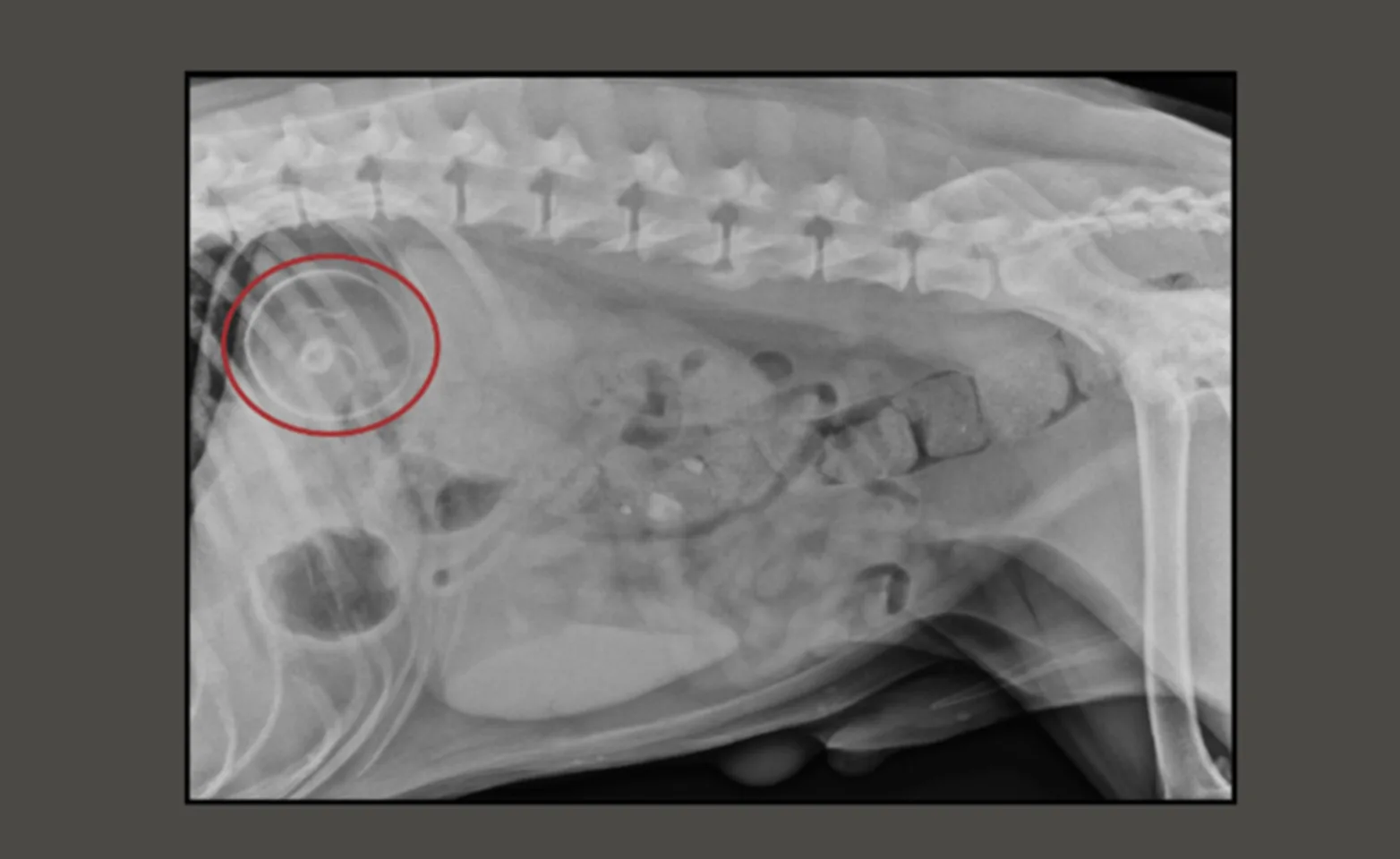
What did Hershey eat?
Hershey came in after stealing and swallowing a squeaky toy from the furry friend next door. Radiographs were performed to confirm the location of the toy which was found in his stomach.
In Hershey's case, surgery was not necessary, and vomiting was induced to present the squeak toy. And it sure did…fully intact!
Hershey did great while in our care and was sent home to begin recovering.
